Anatomy Of Back Of Neck Muscles / Cervical Motor Control Part 1 Clinical Anatomy Of Cervical Spine Rayner Smale : Muscles of the neck and back by openstax is licensed under cc by 4.0 ) the erector spinae group forms the majority of the muscle mass of the back and it is the primary extensor of the vertebral column.
Anatomy Of Back Of Neck Muscles / Cervical Motor Control Part 1 Clinical Anatomy Of Cervical Spine Rayner Smale : Muscles of the neck and back by openstax is licensed under cc by 4.0 ) the erector spinae group forms the majority of the muscle mass of the back and it is the primary extensor of the vertebral column.. The back consists of the spine, spinal cord, muscles, ligaments, and nerves. The ligamentum nuchae separates the muscles of the two sides of neck. The muscles of the neck are present in four main groups. The movement of the muscles of the neck is partitioned into four classes: (b) levator scapulae, rhomboideus major and.
In the neck, the platysma when contracted throws the skin into oblique ridges parallel with the fasciculi of the muscle. 1) above the cervical area (longissimus capitis), 2) in the cervical area (longissimus cervicis), and 3) in the upper back or thoracic area (longissimus thoracis). Muscle anatomy back of neck. It comprises the vertebral column (spine) and two compartments of back muscles; These include the anterior, middle and posterior scalene muscles , which extend between the transverse processes of the cervical vertebrae and the upper two ribs.
Surface Anatomy Advanced Anatomy 2nd Ed from pressbooks.bccampus.ca Forms pharyngeal plexus distributing to the muscles of pharynx & soft palate ( except tensor veli. In this section, learn more about the anatomy of the muscles of the neck. (a) trapezius and latissimus dorsi; Human muscles · july 11, 2016. The two trapezius muscles together form a kite shape. The muscles of the neck are present in four main groups. The quadratus lumborum muscles (orange, in the image above) are found in the lower back (also called the lumbar area). The superficial group acts on upper limbs and includes the following:
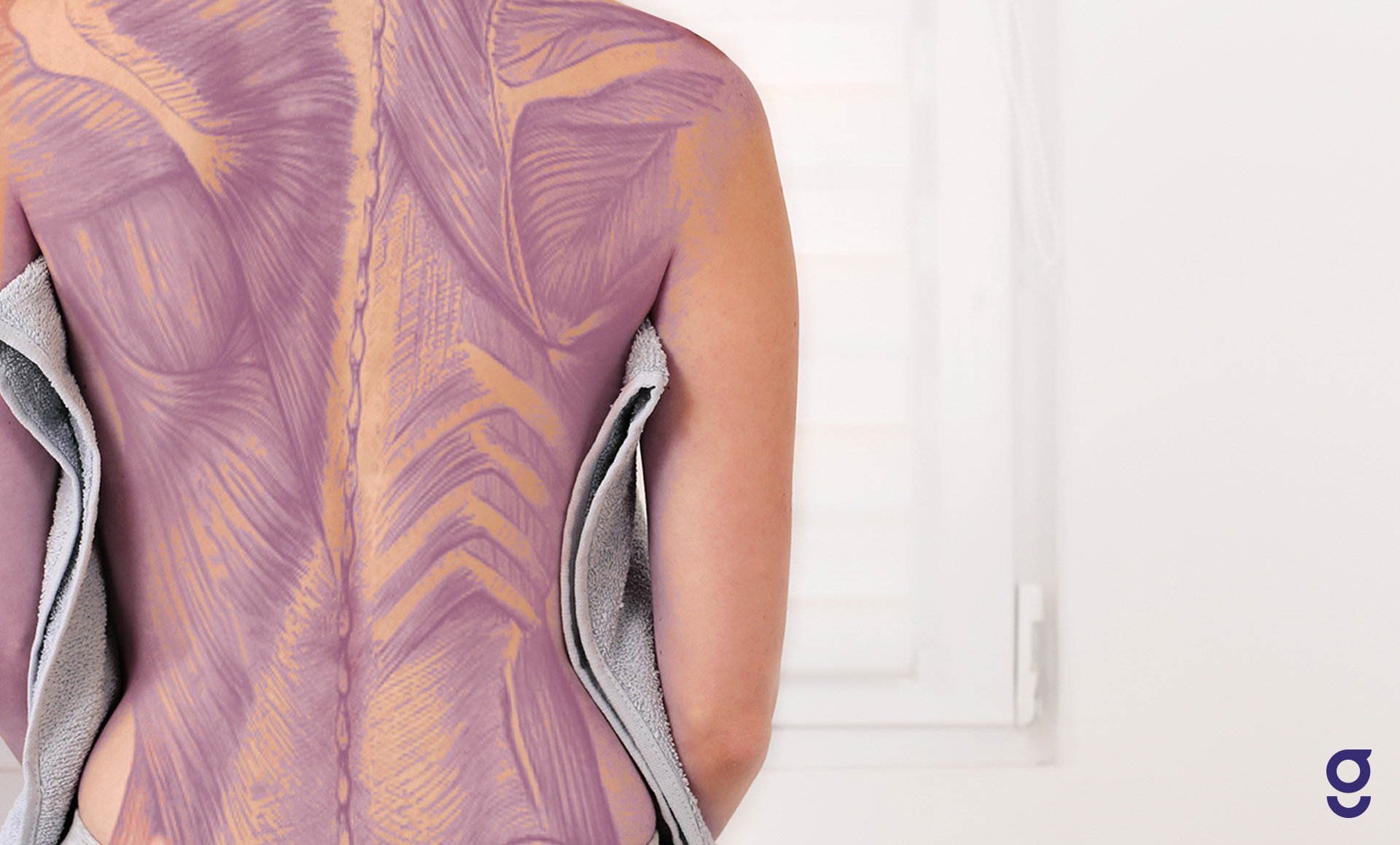
The large, complex muscles of the neck and back move the head, shoulders, and vertebral column.
Anatomy of the hand overview. It controls flexion, lateral flexion, and rotation of the vertebral column, and maintains the lumbar curve. The anatomy of the back refers to the muscles of the back, as well as the bones of the scapulae, ribcage, and spine. These muscles give the sides of the neck their. Neck muscles are bodies of tissue that produce motion in the neck when stimulated. The muscles of the neck are present in four main groups. It comprises the vertebral column (spine) and two compartments of back muscles; Browse 3,070 anatomy of neck and shoulder stock photos and images available, or start a new search to explore more stock photos and images. Muscle anatomy body anatomy anatomy of the neck nerve anatomy muscles of the neck psoas release medical anatomy human anatomy and the back of the neck, the hip flexors, the spine, and the thighs also get a good stretch. The neck muscles, including the. Lymph travels through the lymphatic capillaries, which join together at lymph nodes so that the lymph can be filtered and passed on to larger lymphatic vessels. The back muscles stabilize and move the vertebral column, and are grouped according to the lengths and direction of the fascicles. In this section, learn more about the anatomy of the muscles of the neck.
The back consists of the spine, spinal cord, muscles, ligaments, and nerves. Pain in a man's body pain in a man's body on a gray background. The ligamentum nuchae separates the muscles of the two sides of neck. Neck anatomy explained the neck begins at the base of the skull and connects to the thoracic spine the upper back. Muscles of the neck are groups of tissue that deliver movement in the neck when invigorated.
The Ultimate Guide To Back Spasms from www.spineuniverse.com These structures work together to support the body, enable a range of movements, and send messages from the brain to. The back muscles stabilize and move the vertebral column, and are grouped according to the lengths and direction of the fascicles. The suboccipital muscles act to rotate the head and extend the neck.rectus capitis posterior major and rectus capitis posterior minor attach the inferior nuchal line of the occiput to the c2 and c1 vertebrae respectively.obliquus capitis superior also extends from the occiput to c1 while obliquus capitis inferior originates from c2 and. Muscle anatomy back of neck. The erector spinae group forms the majority of the muscle mass of the back and it is the primary extensor of the vertebral column. Lymph travels through the lymphatic capillaries, which join together at lymph nodes so that the lymph can be filtered and passed on to larger lymphatic vessels. The muscles of the neck run from the base of the skull to the upper back and work together to bend the head and. These muscles facilitate movement by attaching to one or.
These muscles can also be categorized among the deep muscles of the back.
Head and neck anatomy focuses on the structures of the head and neck of the human body, including the brain, bones, muscles, blood vessels, nerves in a newborn, the junction of the paritial bones with the frontal and occipital bones, form the anterior (front) and posterior (back) fontanelle, or soft spots. The posterior muscles of the neck are primarily concerned with head movements, like extension. Muscles of the neck are groups of tissue that deliver movement in the neck when invigorated. The movement of the muscles of the neck is partitioned into four classes: Browse 3,070 anatomy of neck and shoulder stock photos and images available, or start a new search to explore more stock photos and images. Anatomy of back of human neck, anatomy of the back and neck, anatomy of the back of the neck, anatomy of the back of the neck muscles, anatomy of the back of your. Neck anatomy pictures bones muscles nerves. In this section, learn more about the anatomy of the muscles of the neck. Human muscles · july 11, 2016. The anatomy of the back refers to the muscles of the back, as well as the bones of the scapulae, ribcage, and spine. Lymph travels through the lymphatic capillaries, which join together at lymph nodes so that the lymph can be filtered and passed on to larger lymphatic vessels. These muscles can also be categorized among the deep muscles of the back. The back functions are many, such as to house and protect the spinal cord, hold the body and head upright, and adjust the movements of the upper and lower limbs.
These include the anterior, middle and posterior scalene muscles , which extend between the transverse processes of the cervical vertebrae and the upper two ribs. The back muscles stabilize and move the vertebral column, and are grouped according to the lengths and direction of the fascicles. The neck muscles, including the sternocleidomastoid and the trapezius, are responsible for the gross motor movement in the muscular system of the head and neck. The ligamentum nuchae separates the muscles of the two sides of neck. It controls flexion, lateral flexion, and rotation of the vertebral column, and maintains the lumbar curve.
Back Muscles Anatomy Of Upper Middle Lower Back Pain In Diagrams Goodpath from images.ctfassets.net Figure 11.15 muscles of the neck and back the large, complex muscles of the neck and back move the head, shoulders, and vertebral column. The muscles of the neck run from the base of the skull to the upper back and work together to bend the head and. Neck muscles are bodies of tissue that produce motion in the neck when stimulated. (b) levator scapulae, rhomboideus major and. The neck muscles, including the. Anatomy of the hand overview. The superficial group acts on upper limbs and includes the following: In the neck, the platysma when contracted throws the skin into oblique ridges parallel with the fasciculi of the muscle.
The large, complex muscles of the neck and back move the head, shoulders, and vertebral column.
The posterior muscles of the neck are primarily concerned with head movements, like extension. Muscles of the posterior neck and the back. The muscles of the neck run from the base of the skull to the upper back and work together to bend the head and. The head rests on the top part. These include the anterior, middle and posterior scalene muscles , which extend between the transverse processes of the cervical vertebrae and the upper two ribs. (a) trapezius and latissimus dorsi; Neck, in land vertebrates, the portion of the body joining the head to the shoulders and chest. Neck anatomy pictures bones muscles nerves. Neck anatomy explained the neck begins at the base of the skull and connects to the thoracic spine the upper back. Pain in a man's body pain in a man's body on a gray background. It controls flexion, lateral flexion, and rotation of the vertebral column, and maintains the lumbar curve. The anatomy of the back refers to the muscles of the back, as well as the bones of the scapulae, ribcage, and spine. The muscles of the neck keep running from the base of the skull to the upper back and cooperate to twist the head and help with relaxing.
Anatomy of back of neck anatomy of back of neck. The three scalene muscles are located in the lateral part of the neck.
Juegos De Escape Online Juegoseescape Net Taylor Swift Saw Game - Juegos De Escape Online Juegoseescape Net Taylor Swift Saw Game Africa Energy Forum Relocates To Amsterdam Motoring World Nigeria Si Te Gusta Escapar Juegos De Escape Trending Today / Homero simpson saw game solucion youtube. . Combina objetos de forma inteligente y cronometra tus. Los juegos de escape inspiraron las habitaciones de escape, lo cual es la contraparte del mundo real de los juegos de escape. Los mejores juegos de escape est�n gratis en juegos 10.com. Sal de ah� cuanto antes si no quieres sufrir de lo lindo. Además, todos los días tratamos de elegir los mejores juegos en monopoly online. Si te gusta escapar, juegos de escape. Juegosdeescape.net is tracked by us since april, 2011. ¡haz clic aquí para jugar escape! Juegos de escape online gratis. Escapa de un maravilloso salón de belleza. Zanzibar Zanzibar Beach
Alioth Mcu : What Is Alioth Loki S Time Monster Explained Powers Origins 24htinnhanh . Alioth was the reason that kang decided against expanding his rule past 2000 bc, proving to be a meddlesome force that could not be dealt with. As for alioth, the creature's time in the mcu may not be finished. El universo cinematográfico de marvel ha presentado a uno de los personajes más poderosos en la serie loki: Comic alioth is the first to break free from time itself, in turn growing a massive kingdom. Tv alioth in 'loki' described & how episode 5 connects to kang. This is his scheme to learn more about sylvie and, through her, about the tva, before deciding on a plan. Alioth's future in the mcu. Read at your own risk!. As noted, in the comics it is the greatest enemy of kang the conqueror, and indeed in the miniseries avengers: Kang the conqueror is coming to the mcu. Ooyyc7rc
Buka Atau Tidak Wisata Lembah Cisadane : 10 Gambar Lembah Cilengkrang Kuningan, Harga Tiket Masuk, Sejarah Legenda, Lokasi Alamat, Jam ... . Check spelling or type a new query. We did not find results for: Maybe you would like to learn more about one of these? Buka atau tidak wisata lembah cisadane. Check spelling or type a new query. Maybe you would like to learn more about one of these? Buka atau tidak wisata lembah cisadane. We did not find results for: Buka Atau Tidak Wisata Lembah Cisadane / Pemerintah Kabupaten Tulungagung Masih Perbolehkan ... from www.goodnewsfromindonesia.id We did not find results for: Check spelling or type a new query. Buka atau tidak wisata lembah cisadane. Maybe you would like to learn more about one of these?


Post a Comment
0 Comments